
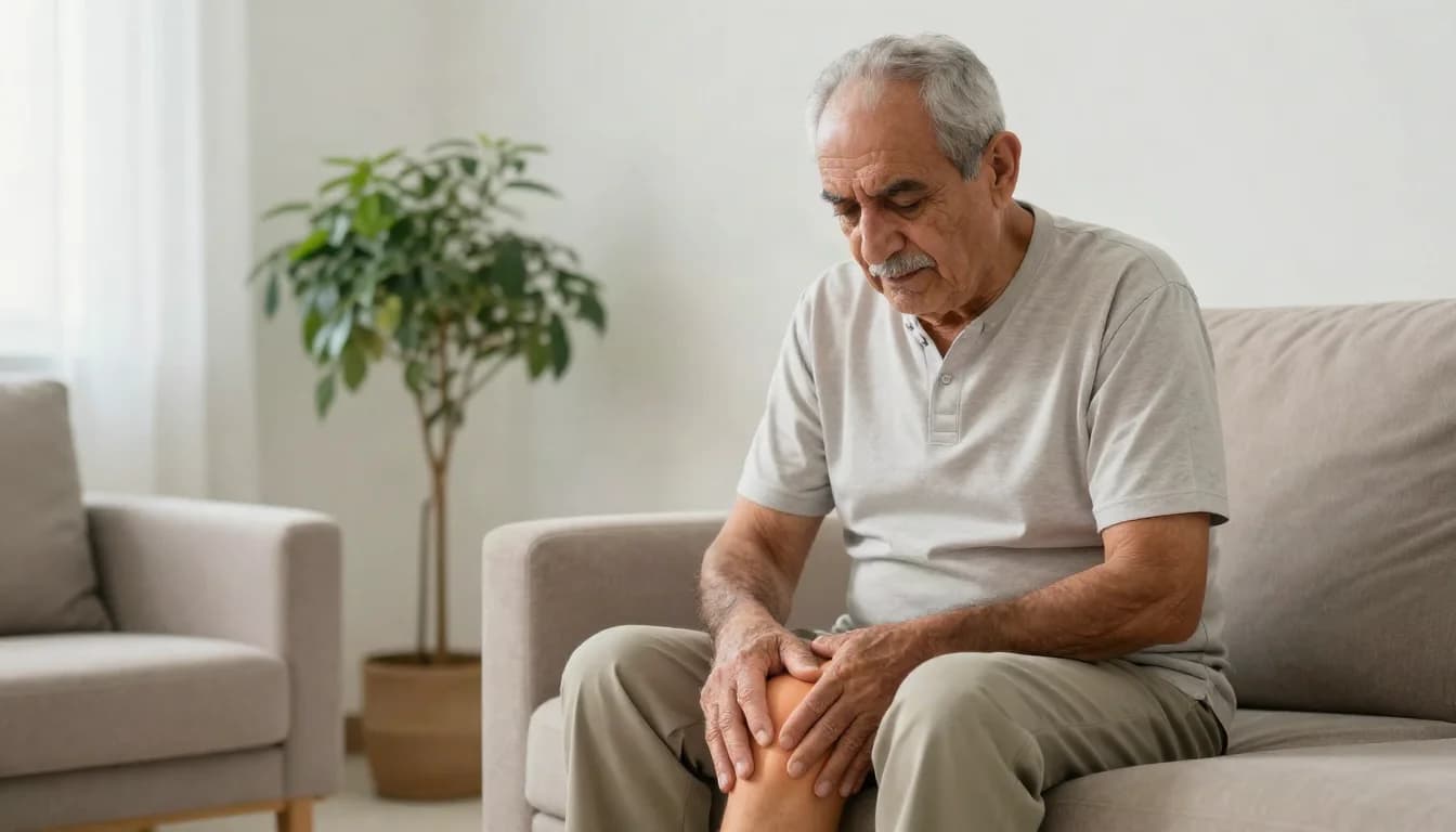
Spinal stenosis is a narrowing of the space through which the spinal cord and nerve roots travel inside the spine, or of the openings where nerves exit between the vertebrae, and it is this narrowing that produces symptoms by pressing on neural structures. The most common causes are degenerative changes that accumulate with age: enlargement of the facet joints, thickening of the ligamentum flavum, bulging discs, and slippage of one vertebra on another. In the lower back the classic picture is neurogenic claudication: pain, heaviness, numbness or burning in the legs that appears after walking a certain distance or standing for a while, and eases on sitting, leaning forward or resting on a trolley, so the distance a person can walk shrinks over time. In the neck, stenosis may instead cause myelopathy: heavy hands, loss of finger dexterity, difficulty with shirt buttons and unsteadiness on the feet. That picture calls for prompt assessment rather than watchful waiting. Treatment usually starts on the non-surgical track: physiotherapy centred on flexion-based positions that functionally open the canal, core strength and general fitness; activity modification; weight reduction; medication prescribed by the doctor to control pain; and image-guided epidural injection in selected cases. When that fails, or when walking is severely limited or there is clear neurological weakness, decompression surgery is considered, removing the compressing bone and ligament through a small incision or endoscope in suitable cases, with fusion added if there is instability or slippage. The limits matter. Exercise and injections relieve symptoms but do not anatomically widen the canal; only surgery does that. An injection is a temporary measure intended to calm inflammation around the nerve root and can help identify the source of pain, but it is not a permanent treatment and cannot be repeated indefinitely. Surgery is generally better at improving leg pain and walking capacity than at removing chronic low back pain, it does not fully reverse nerve damage that has persisted for a long time, and it does not stop degeneration at the remaining levels. The word stenosis in an MRI report is also not by itself a reason for surgery. The decision rests on whether your symptoms match the site of narrowing and the findings on neurological examination, and your candidacy is determined by clinical assessment, not by reading a report or a web page.
Procedure steps
- 1
History taking and distinguishing claudication
The doctor asks how far you walk before pain starts and what relieves or aggravates it, distinguishes neurogenic claudication from arterial claudication caused by poor circulation, and examines foot pulses, power, sensation, reflexes, gait and balance.
- 2
Appropriate imaging
MRI is the most accurate way to show the degree and levels of narrowing and the state of the nerve roots. Standing, flexion and extension X-rays are added to assess instability, CT is used when MRI is not possible, and nerve conduction studies help when peripheral neuropathy confuses the picture.
- 3
A structured non-surgical programme
A physiotherapy programme using flexion-based positions, core strengthening, a stationary bicycle and graded walking, along with activity modification, weight reduction and medication prescribed by the doctor. Response is measured against practical markers: walking distance, sleep quality, and the ability to work, pray and shop.
- 4
Image-guided injection in selected cases
An epidural or nerve-root injection performed under image guidance to calm inflammation around the compressed root, used to break the pain cycle and make rehabilitation possible, or to confirm which level is responsible. Its effect varies between patients and is temporary, and repetition is limited by rules your doctor sets.
- 5
Decompression surgery and follow-up
Under general anaesthesia the compressing enlarged bone, thickened ligament and disc tissue are removed, through a small incision or endoscope in suitable cases, with fusion added if there is instability. Walking begins the same or the next day, rehabilitation progresses over weeks, and neurological symptoms are followed.
Before the procedure
Before your visit, record how far you walk before pain begins and what relieves it; this single detail is worth as much as a full report. Bring your MRI discs, films and written reports, not just summaries. Tell the doctor about chronic conditions, particularly diabetes, blood pressure and peripheral arterial disease, and about every medicine you take including blood thinners, aspirin and anti-inflammatories, and do not stop anything on your own. If you are a candidate for an image-guided injection, mention any allergy to contrast or medication and any active infection or fever, and arrange for someone to drive you home afterwards rather than driving yourself. If surgery is planned, stop smoking, bring blood sugar under control, complete the requested tests and follow fasting instructions.
After the procedure
After an image-guided injection, rest relatively for the rest of the day and then return to activity gradually. Expect that pain sometimes flares briefly before it improves, and monitor your blood sugar if you are diabetic, because steroid can raise it for a few days. After decompression surgery, take short frequent walks from day one, avoid prolonged sitting, heavy lifting and repeated bending for as long as your surgeon specifies, keep the wound clean and dry, and commit to the physiotherapy programme, because the result depends on it as much as on the operation. **Go to the emergency department immediately for progressive leg weakness or a dropping foot, numbness between the thighs and around the anus, urinary retention, or leakage of urine or stool — these are cauda equina symptoms and are treated as an emergency.** Contact your doctor quickly for fever or chills, clear fluid leaking from the wound with a headache that worsens on standing and eases on lying down, pain and swelling in the leg, or new severe back pain.
Expected duration
An assessment visit usually takes 20 to 40 minutes, an image-guided injection usually 15 to 30 minutes, and decompression surgery usually one to three hours.
Our directory of Spinal canal stenosis treatment doctors in Jordan is growing
There are no matching published profiles in this directory yet. ClinicsJo is expanding its coverage and working to welcome more Spinal canal stenosis treatment doctors in Jordan. Check this page regularly for newly added profiles, practice details and available contact options.
Browse all doctorsBrowse Spine profilesFinding Spinal canal stenosis treatment services in Jordan
Which doctors are listed for Spinal canal stenosis treatment in Jordan?
There are currently no doctor profiles linked to Spinal canal stenosis treatment in this directory. You can browse the related specialty or the wider doctors directory. This does not mean that the service is unavailable in Jordan.
How can I find an appointment for Spinal canal stenosis treatment?
Start with an available doctor's profile and use the contact or booking options shown there. Confirm the service, practice location and appointment time with the clinic. If this list is empty, broaden your search using the directory links on this page.
What does Spinal canal stenosis treatment cost in Jordan?
Ask the practice for the current price and what it includes. A consultation fee shown on a profile is not necessarily the price of a procedure, tests or follow-up. Confirm any additional charges and insurance arrangements before your visit.