

Jordan Eye Center
Dr Salem Ahmad Abu Al Ghanam’s ophthalmic work centres on glaucoma and elevated eye pressure. His areas of care include Ahmed valve implantation and laser and u…
Hours not listedInsurance not specified8 views
Trusted medical platform in Jordan
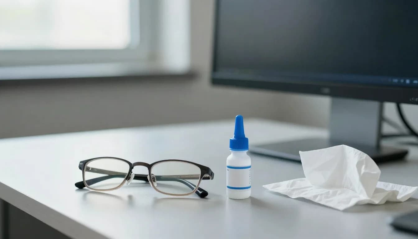
Glaucoma, often called raised eye pressure, is a group of diseases that progressively damage the optic nerve, usually (though not always) alongside raised pressure of the fluid inside the eye. Treatment aims to lower eye pressure to an individual target, because pressure is the only factor we know how to control in order to slow nerve damage. Three tools are available: drops, laser, and surgery when the first two are not enough. The most dangerous feature of this disease is that **it steals sight with no early symptoms**. The common chronic type causes no pain, no redness and no early blurring; loss begins at the edges of the visual field where nobody notices it, and the better eye masks the deficit of the other, so vision seems normal until advanced stages. Glaucoma is therefore found by examination, not by complaint. Anyone with an affected relative, over forty, on long-term steroids, with diabetes or with high myopia needs periodic eye-pressure and optic-nerve checks even while feeling perfectly well. Assessment includes measuring eye pressure, examining and imaging the optic nerve with optical coherence tomography to quantify nerve-fibre thickness, visual field testing, corneal thickness measurement, and examination of the drainage angle to classify the type as open or closed angle. These tests are repeated over time, because treatment decisions rest on the trend rather than on a single pressure reading. A target pressure is then set for each eye according to the extent and speed of damage. The limits must be stated plainly: **optic nerve damage cannot be reversed**, and no treatment restores a portion of visual field that has been lost. Drops, laser and surgery all exist **to preserve what remains, not to improve vision**, so do not expect to see better after starting a drop. This creates the most dangerous practical problem in glaucoma care: because the patient feels neither benefit nor harm, drops are abandoned after a few weeks, pressure returns and damage continues silently. **Daily, on-time use of the drop is the treatment itself.** Do not stop it, do not stretch the intervals, do not wait for symptoms, and if the bottle runs out before your appointment, contact your doctor rather than simply stopping. To correct a widespread myth: glaucoma is not a condition removed once and for all by a single operation, and it is not the same as cataract, which is treated by replacing the lens. Glaucoma is chronic and needs lifelong monitoring even after successful surgery. Eligibility for any plan is determined by clinical examination with serial visual fields and nerve imaging.
Eye pressure measurement, optic nerve examination with OCT imaging, visual field testing, corneal thickness measurement and gonioscopy of the drainage angle. The glaucoma is classified as open angle, angle closure, secondary or normal-tension, because each type follows a different treatment path, and medications and comorbidities are documented.
A numerical target is chosen according to the extent and rate of nerve damage, the patient's age, corneal thickness and family history. There is no single target that suits everyone, and it is revised downward if the visual field or nerve-fibre imaging shows progression despite reaching the first target.
One drop is usually started and pressure rechecked after a few weeks to gauge response, with a second agent added or a substitution made as needed. The patient is trained in correct technique: a single drop, gentle eye closure without squeezing, pressing the inner corner for a few minutes to reduce systemic absorption, five minutes between two different drops, and tying each dose to a fixed alarm.
In open-angle disease, laser trabeculoplasty may be used as a first-line or supporting option, while in a narrow angle a laser opening in the peripheral iris prevents an acute closure attack. The session is done in clinic under anaesthetic drops, does not remove the need for monitoring, and some patients will need drops again later.
Pressure, visual fields and nerve imaging are repeated at intervals set by the ophthalmologist so that progression is caught early; this comparison over time is the essence of follow-up. If damage continues despite adherence and laser, if drops are not tolerated, or if adherence is not achievable, surgery is discussed with a frank explanation of its aims and limits.
Bring every drop and medicine you use in its own bottle rather than reciting names from memory, and tell the doctor if you use corticosteroids in any form — drops, inhaler, tablets or ointment — because they can raise eye pressure. Mention asthma, a slow heart rate, kidney stones or drug allergies, as these rule out certain classes of drops. Bring previous visual field and nerve imaging reports if they were done elsewhere, because comparison with the old test matters more than a new test alone. Expect your pupils to be dilated, so do not drive yourself and do not plan detailed work afterwards, and bring sunglasses. Do not skip your drop on the day of the examination unless your doctor explicitly asks you to.
Use the drop on time every day even if you feel no difference, because the goal is to protect what remains rather than to improve vision. Do not stop it or reduce the frequency on your own, do not share it, and request a repeat prescription before the bottle runs out. Press the inner corner of the eye for a few minutes after instilling to reduce systemic absorption, and leave five minutes between two different drops. Report persistent redness, itching, a change in iris colour, lash growth, breathlessness or a slow pulse. Keep your visual field and imaging appointments even while you feel well. **Go to emergency care immediately if** you develop severe eye pain with headache, nausea or vomiting, halos around lights, marked redness and sudden blurring, as these suggest an acute angle-closure attack that threatens sight within hours. **Seek prompt review** for any sudden drop in vision, a new black patch, or pain that does not settle.
A full assessment visit usually takes 45 to 90 minutes depending on the tests; drop treatment is daily and long term, with follow-up typically every 3 to 6 months.
Dr Salem Ahmad Abu Al Ghanam’s ophthalmic work centres on glaucoma and elevated eye pressure. His areas of care include Ahmed valve implantation and laser and u…
There are currently 1 doctor profiles linked to Glaucoma Treatment on ClinicsJo. Review the listed services and contact the practice to confirm availability with the doctor and branch you choose.
Start with an available doctor's profile and use the contact or booking options shown there. Confirm the service, practice location and appointment time with the clinic. If this list is empty, broaden your search using the directory links on this page.
Ask the practice for the current price and what it includes. A consultation fee shown on a profile is not necessarily the price of a procedure, tests or follow-up. Confirm any additional charges and insurance arrangements before your visit.