
Al Shamaileh Cardiology Clinic
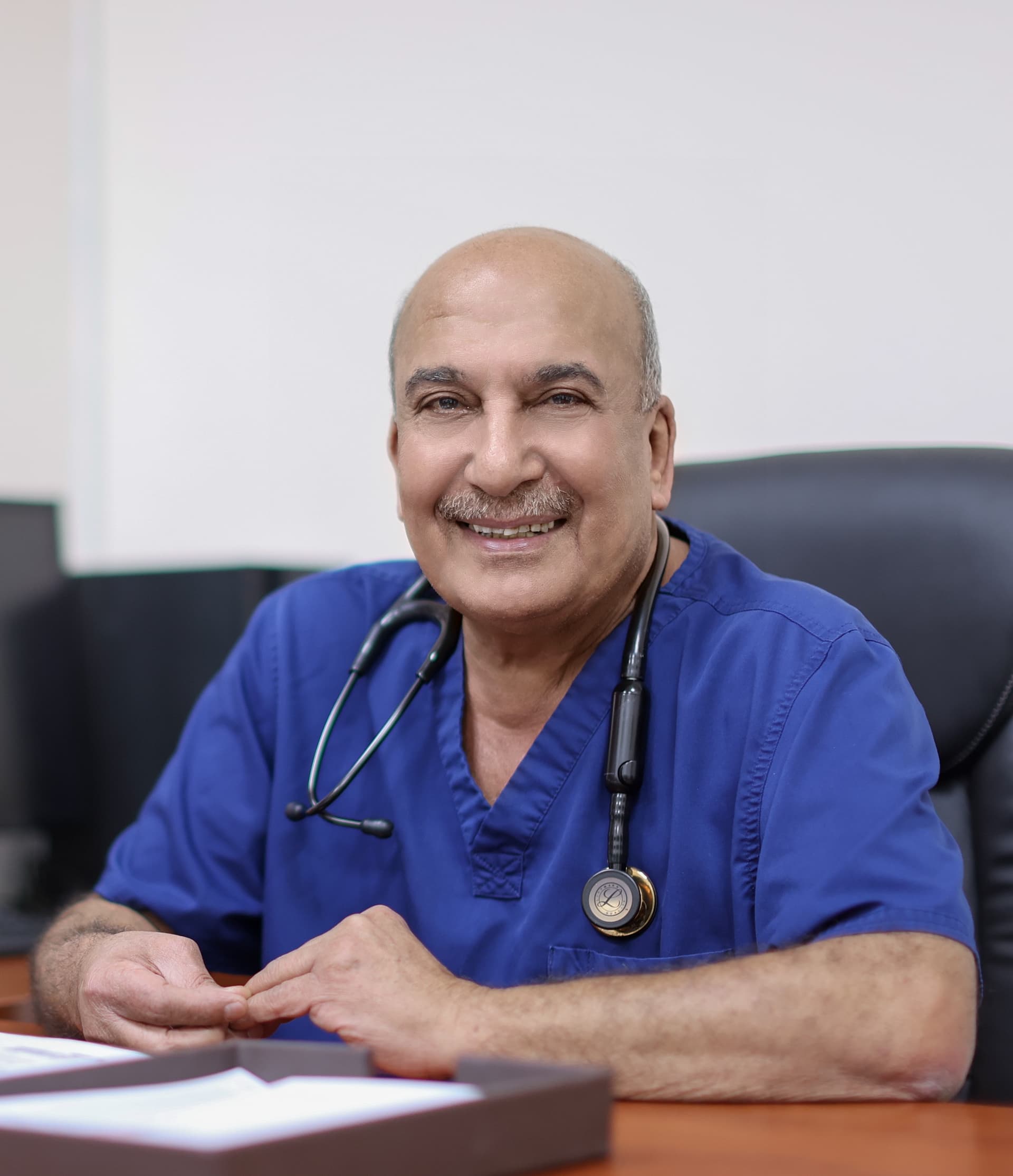
Dr. Qasem Al Shamaileh is a consultant in cardiology and interventional cardiology. His practice includes coronary and valve assessment, selection of appropriat…
Hours not listedInsurance not specified12 views
Trusted medical platform in Jordan

إغلاق الفتحة بين الأُذينين
Transcatheter closure of an atrial septal defect seals a congenital hole in the wall between the two upper heart chambers using a small mesh occluder device. The device is advanced folded inside a catheter from a vein in the upper thigh to the heart, then opened on both sides of the septum as two discs that grip the defect from front and back. The chest is not opened and the heart is not stopped; the procedure is guided by X-ray and by echocardiography throughout. An atrial septal defect lets part of the blood return to the right side and the lungs instead of leaving for the body, so the right ventricle carries extra volume for many years. Symptoms may therefore be absent in childhood and begin in early adulthood or later: breathlessness on exertion, easy fatigue, palpitations or rhythm disturbance, and repeated chest infections in children. Accepted indications for closure are enlargement of the right side of the heart, increased flow to the lungs, or symptoms — not simply the presence of a small hole on an echocardiogram report. The limits are clear: **catheter closure is not suitable for every type of defect.** The mid-septal type with adequate rims around the hole is the appropriate candidate; other types lying near the valves or near the vein entrances, very large defects, defects with deficient rims, and some multiple defects are better treated by open-heart surgery. The procedure also **does not treat pulmonary hypertension once it has become advanced and fixed**, does not guarantee that rhythm disturbance will disappear in older patients who have lived with the defect, and does not return the right ventricle to normal size immediately — improvement is gradual over months. Suitability is determined by clinical examination and detailed echocardiography measuring the defect and its rims, not by reading. The device is gradually covered by the heart's own tissue over months and becomes part of the septum, which is why antiplatelet medication is prescribed for a period and temporary caution is advised regarding vigorous and contact sports. **See a doctor immediately for fainting, new persistent palpitations, severe chest pain, increasing breathlessness, sudden weakness of the face or a limb or slurred speech, or fever with chills.**
Beforehand a detailed echocardiogram — often a transoesophageal study — measures the diameter of the defect and the thickness of its rims, confirms its type, checks the veins and valves, and verifies that pulmonary pressure permits closure. The device size is chosen from these measurements.
The procedure is done under general anaesthesia in children and often under local anaesthesia with sedation in adults. A sheath is placed in a vein in the upper thigh — a vein rather than an artery, which makes the access site easier to manage — and the catheter is advanced through it into the right atrium.
The catheter is passed from the right atrium to the left through the defect itself, and right-sided pressures, pulmonary artery pressure and flow ratios are measured. These numbers determine whether closure will genuinely help or whether pulmonary hypertension makes it inadvisable.
The folded device is advanced into the left atrium where its first disc opens, then it is drawn back gently against the septum and the second disc is opened on the right side. It is released from its delivery cable only after echocardiography confirms it is stable and is not obstructing a valve or a vein.
Bleeding at the venous site is controlled with pressure and a dressing, and pulse, ECG and the access site are monitored for several hours. An echocardiogram and ECG confirm device position with no significant residual shunt or rhythm disturbance, and discharge is usually the following day.
Tell your doctor about every medicine you take, especially anticoagulants and antiplatelet drugs, since some are adjusted before the procedure on the doctor's instruction alone; never stop a medicine yourself. Report your kidney function if you have a recent result, and any previous reaction to X-ray contrast, iodine or metals. Raise any active infection before the date — gum infection, a dental abscess, or a urinary or skin infection — because implanting a device inside the heart while an infection is present is hazardous and is better postponed and treated first; a dental check beforehand is advised. Fast for the period the team specifies, say if you are or might be pregnant, bring previous echocardiogram reports, and arrange a companion and an overnight stay. For children, follow fasting instructions carefully and bring something comforting.
Look after the groin puncture site: keep the dressing clean and dry, and avoid lifting and strenuous effort in the first days as the team advises. **Go to the emergency department immediately for: increasing swelling, bleeding or severe pain at the access site; a cold, blue or numb leg; fainting or new persistent palpitations; severe chest pain; increasing breathlessness; sudden weakness of the face or a limb or slurred speech; or fever with chills and night sweats — which may indicate infection inside the heart, so do not start an antibiotic yourself before being examined.** **Do not stop or alter the antiplatelet medicine prescribed after your device on your own, not even for a tooth extraction; the doctor who implanted it decides.** Tell every dentist and surgeon that you have an intracardiac closure device, follow the infection-prevention advice before dental work in the early months, keep your echocardiogram and follow-up appointments, and avoid contact sports for the period your doctor specifies.
Usually 45 to 90 minutes, a little longer when the defect anatomy is complex or transoesophageal echocardiography is required, followed by several hours of monitoring, most often a single overnight stay, and an echocardiogram to confirm device position before discharge.
Dr. Qasem Al Shamaileh is a consultant in cardiology and interventional cardiology. His practice includes coronary and valve assessment, selection of appropriat…
There are currently 1 doctor profiles linked to Atrial septal defect (ASD) closure on ClinicsJo. Review the listed services and contact the practice to confirm availability with the doctor and branch you choose.
Start with an available doctor's profile and use the contact or booking options shown there. Confirm the service, practice location and appointment time with the clinic. If this list is empty, broaden your search using the directory links on this page.
Ask the practice for the current price and what it includes. A consultation fee shown on a profile is not necessarily the price of a procedure, tests or follow-up. Confirm any additional charges and insurance arrangements before your visit.