
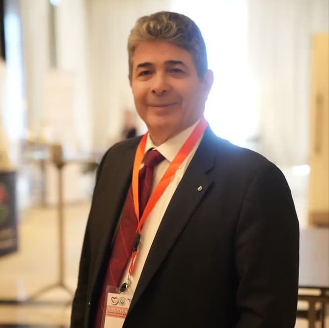
CardioCare Clinic – Dr Marwan AlNimri
Dr Marwan Adeeb AlNimri is a consultant interventional cardiologist with Jordanian boards in internal medicine and cardiology and international professional fel…
Hours not listedInsurance not specified5 views
Trusted medical platform in Jordan

An echocardiogram is an ultrasound scan that shows the heart while it beats: the size of the chambers, the thickness of the muscle and how strongly it moves, the shape and motion of the valves and the direction of blood flowing through them, how much the left ventricle ejects with each beat, and the sac around the heart and whether fluid has collected in it. The test uses no radiation, causes no pain, and in most cases needs no needle. It is requested for breathlessness, leg swelling, a heart murmur, palpitations or fainting; to follow long-standing high blood pressure; to assess a weakened heart muscle; after a heart attack; before major surgery; to monitor a repaired or prosthetic valve; and when cardiac symptoms appear in pregnancy. Both the indication and the interpretation are determined by clinical assessment, not by reading a report in isolation. There are several types. A transthoracic echo is the commonest and is performed with the probe on the skin of the chest. A transoesophageal echo is done with sedation and fasting, passing a slim probe through the throat for sharper images of the valves and the left atrium. A stress echo images the heart before and after exertion to assess exercise-related symptoms. Contrast agents are used in selected cases. The scan is done while you lie on your left side with gel on the chest and simple ECG electrodes attached. You may be asked to hold your breath briefly, and you will hear Doppler sounds, which are normal and do not signal a problem. The limits are among the most important things to understand. **An echocardiogram does not show narrowing of the coronary arteries.** A normal echo does not rule out coronary artery disease and does not make unexplained chest pain safe, which is why other tests such as an ECG, a stress test or coronary imaging are requested. It does not capture intermittent rhythm disturbances that come and go — those need an ECG during symptoms or prolonged monitoring — and it measures neither cholesterol nor blood pressure. Image quality can be limited by excess weight, lung disease or chest wall deformity, and a single number in the report, such as the ejection fraction, is never read alone but alongside your symptoms and your other results.
The type is chosen according to the clinical question: a transthoracic echo for a general assessment of heart function and valves, a transoesophageal study for sharper images in specific situations, or a stress echo when symptoms are related to exertion. Ask your doctor exactly what your scan is looking for.
The chest is exposed, simple ECG electrodes are attached and you lie on your left side. Water-based gel is applied to the skin so the sound waves can pass, and the room light is dimmed so the screen can be seen. A standard scan needs no fasting, no injection and no interruption of medicines.
The probe is moved over defined positions on the chest, at the base of the neck and below the breastbone, clips of the chambers and valves are recorded, and dimensions and blood velocities are measured with Doppler. You may be asked to hold your breath briefly, and the pulsing sounds you hear are a normal part of the study.
A transoesophageal echo requires fasting, sedation and an escort, with local anaesthetic to the throat. A stress echo combines resting images with images taken after exercise on a treadmill or after a pharmacological stimulus, with the ECG and blood pressure monitored throughout.
A report describes chamber sizes, muscle and valve function and estimated pressures. It is read together with your symptoms, your ECG and your blood tests, and from it the team decides whether further tests are needed, whether the medication plan changes, and when a follow-up echo is due.
A transthoracic echo needs no fasting and no special preparation: eat and drink normally, wear clothing that opens at the front, and do not apply creams or oils to the chest on the day. Bring your medication list, previous reports and any old echo or ECG, because comparison with an earlier study changes interpretation considerably. **Do not stop your cardiac medicines, antiplatelets or anticoagulants before the scan on your own — an echo does not require stopping them at all, and any interruption before any other procedure, including a tooth extraction or dental cleaning, is a decision for your cardiologist, not for you and not for the dentist alone.** For a transoesophageal study, follow the fasting instructions, arrange an escort because you will be sedated, and tell the team about swallowing difficulty, oesophageal problems or loose dentures. For a stress echo, wear trainers and ask about the timing of your rate-controlling medicines.
After a transthoracic echo there are no restrictions at all: wipe off the gel and return to your work, your food and your medicines exactly as before. After a transoesophageal study, do not eat or drink until normal swallowing sensation has returned so you do not choke, expect a mildly sore throat that settles within a day, do not drive or make important decisions on the day of sedation, and have someone accompany you home. **Go to the emergency department immediately for chest pain or pressure, breathlessness, fainting or severe dizziness, or a racing heartbeat with dizziness — do not wait for your results appointment.** **After a transoesophageal echo, seek help urgently for severe chest or abdominal pain, increasing pain or difficulty swallowing, vomiting blood or high fever**, as these can indicate a rare oesophageal injury needing prompt assessment. Never adjust your medication because of a number in the report; the report is read together with your doctor.
A transthoracic echo usually takes 20 to 45 minutes, a transoesophageal study about 20 to 30 minutes plus an hour or more of recovery after sedation, and a stress echo typically 45 minutes to an hour.
Dr Marwan Adeeb AlNimri is a consultant interventional cardiologist with Jordanian boards in internal medicine and cardiology and international professional fel…
There are currently 1 doctor profiles linked to Echocardiography on ClinicsJo. Review the listed services and contact the practice to confirm availability with the doctor and branch you choose.
Start with an available doctor's profile and use the contact or booking options shown there. Confirm the service, practice location and appointment time with the clinic. If this list is empty, broaden your search using the directory links on this page.
Ask the practice for the current price and what it includes. A consultation fee shown on a profile is not necessarily the price of a procedure, tests or follow-up. Confirm any additional charges and insurance arrangements before your visit.