Upper gastrointestinal endoscopy (oesophagogastroduodenoscopy) is a diagnostic and therapeutic procedure in which a thin flexible tube with a camera and light at its tip is passed through the mouth into the oesophagus, stomach and the start of the small intestine, letting the physician see the mucosal surface directly rather than inferring from symptoms or images.
What makes it indispensable is that it is not only looking: within the same session **biopsies can be taken** for histology, Helicobacter pylori tested for, bleeding stopped, a stricture dilated, and a foreign body or polyps removed. It therefore often ends a long debate about the cause of persistent symptoms.
It is usually requested for heartburn or reflux not responding to treatment, persistent upper abdominal pain, difficulty or pain on swallowing, repeated vomiting, or **features requiring urgency**: vomiting blood or black stools, unexplained iron-deficiency anaemia, unexplained weight loss, or progressive difficulty swallowing. Those last ones are not managed with antacids and waiting.
It is most often performed under intravenous sedation that leaves the patient unaware of most of the procedure or comfortable, and takes a few minutes. What worries patients most — choking — does not usually occur, because the endoscope passes into the oesophagus and not the airway, and breathing continues normally.
Preparation is simple but not negotiable: **the specified fasting is essential, because food in the stomach both blocks the view and raises the risk of aspiration into the lungs**. The team must also be told about blood thinners, diabetes, and cardiac and valve disease, as these change preparation.
1 specialists
Procedure steps
1
Establishing the indication and preparing
Symptoms, urgency features and medications are reviewed, the required fasting specified, and a plan made for blood thinners and diabetes drugs in coordination with the treating physician.
2
Intravenous sedation
Intravenous access is placed and sedation given with monitoring of pulse, oxygen and blood pressure; a topical throat spray may reduce the gag reflex.
3
Passing the scope and examination
The scope is passed through the mouth into the oesophagus, stomach and proximal small intestine; a little air distends the wall so the mucosa can be examined carefully and imaged.
4
Biopsies and intervention when needed
Biopsies are taken for histology and H. pylori testing, and bleeding may be stopped, a stricture dilated, or a polyp or foreign body removed in the same session.
5
Recovery and reporting
The patient is monitored until sedation wears off and the findings are explained; biopsy results follow later and the final treatment plan is built on them.
Before the procedure
Follow the fasting instructions for food and fluids exactly as the team specifies — this is the most important instruction, since food cancels the procedure and raises the risk of pulmonary aspiration. Tell the team about all medications: **blood thinners and antiplatelets** (do not stop them yourself — they are managed by plan), diabetes drugs (some are adjusted on the fasting day), and acid suppressants, which may need stopping for a period if H. pylori testing is the goal. Report cardiac and valve disease, drug allergies and any previous anaesthetic problem. Remove dentures before the procedure, and arrange transport both ways, as sedation prohibits driving.
After the procedure
Do not drive, operate machinery or sign important documents on the day of sedation, and have someone accompany you home. Start with fluids then light food once swallowing sensation has fully returned and the team permits, especially if a throat spray was used. Expect bloating, belching and mild throat soreness for a day or two. If a biopsy was taken or a polyp removed, follow any specific dietary or medication instructions given. Ask when biopsy results are due and who will explain them. **Attend emergency care immediately for severe chest or abdominal pain, vomiting blood or black stools, fever and chills, difficulty swallowing or breathing, or neck swelling or increasing pain on swallowing.**
Expected duration
The examination takes 5–15 minutes, with about one to two hours at the centre for recovery
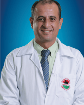
Dr. Ashraf Mohammad Sabri Ali is a specialist in internal medicine, gastroenterology and liver disease at Jordan Hospital. He graduated from Al Mustansiriyah Un…
Which doctors are listed for Esophagogastroscopy in Jordan?
There are currently 1 doctor profiles linked to Esophagogastroscopy on ClinicsJo. Review the listed services and contact the practice to confirm availability with the doctor and branch you choose.
How can I find an appointment for Esophagogastroscopy?
Start with an available doctor's profile and use the contact or booking options shown there. Confirm the service, practice location and appointment time with the clinic. If this list is empty, broaden your search using the directory links on this page.
What does Esophagogastroscopy cost in Jordan?
Ask the practice for the current price and what it includes. A consultation fee shown on a profile is not necessarily the price of a procedure, tests or follow-up. Confirm any additional charges and insurance arrangements before your visit.