
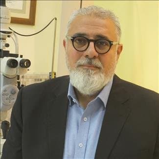
Amman Eye Clinic
Dr. Mohammad Abu Samak is a consultant ophthalmologist with vitreoretinal fellowship training at Henry Ford Hospital in the United States. His clinical work cov…
Hours not listedInsurance not specified5 views
Trusted medical platform in Jordan

Diabetic retinopathy is damage to the tiny blood vessels within the retina caused by long-standing high blood sugar. Vessels become fragile and leak fluid or blood, and abnormal new vessels may grow causing haemorrhage or traction on the retina. It is a leading cause of vision loss in adults — and its most dangerous feature is that it begins **with no symptoms at all**. That point is the heart of the matter: the retina may be damaged to a degree requiring immediate treatment while the patient’s vision is still perfectly good. **Regular dilated retinal examination is therefore not optional for someone with diabetes and is not deferred until a complaint appears** — waiting for vision to change often means losing the window for easy early treatment. Treatment has advanced considerably and depends on disease stage: **intravitreal injections** of anti-vascular-growth agents reduce leakage and swelling at the centre of vision (today the mainstay for diabetic macular oedema, given as multiple doses on a monitoring schedule); **laser** stabilises abnormal vessels and prevents haemorrhage in proliferative stages; and **vitreous surgery** is used for dense non-clearing haemorrhage or traction detaching the retina. But none of these works in isolation from the foundation: **control of blood sugar, blood pressure and lipids is what slows the disease itself**, while eye treatment addresses damage already done. Treating the eye while neglecting glucose invites the disease back. Smoking and kidney disease worsen the condition. In many cases the realistic goal is **preserving remaining vision and preventing deterioration** rather than restoring vision already lost — another reason why early action is everything.
Fundus examination after pupil dilation at intervals set by the ophthalmologist according to diabetes duration and control — performed even with perfect vision, because the disease begins silently.
Macular OCT precisely quantifies swelling, and retinal angiography is used when needed to map leakage and ischaemia and grade the stage.
An anti-vascular-growth agent is injected into the eye under strict asepsis to reduce leakage, repeated in doses on a schedule set by response.
Laser is applied to retinal areas to reduce the drive for abnormal vessel growth and prevent haemorrhage and vision loss, sometimes over more than one session.
Performed for dense non-clearing haemorrhage or traction detaching the retina, removing the vitreous and blood, releasing traction and stabilising the retina.
Bring HbA1c, blood pressure, lipid and kidney results, your diabetes duration and medication list — the eye plan is built on this complete picture. **Arrange transport both ways for examination visits, because pupil dilation blurs vision and increases glare for hours and prohibits driving.** Bring sunglasses. Report previous eye surgery, prior injections or raised eye pressure. If you are pregnant tell your ophthalmologist immediately, as pregnancy can accelerate retinopathy progression and requires closer monitoring.
Do not rub the eye after injection, and use any prescribed drops. Most important: **attend the full injection and review schedule and do not stop because your vision improved** — stopping early is among the commonest causes of oedema returning and losing the gains. Control blood sugar, blood pressure and lipids seriously, as these slow the disease itself, and stop smoking. After injection expect a gritty sensation, mild redness and possibly floaters for a day or two. **Seek urgent care immediately for severe eye pain, sudden visual deterioration, new flashes or a sudden shower of floaters, a shadow or curtain moving across the visual field (which may mean retinal detachment), or increasing redness with discharge and marked light sensitivity after injection.**
Examination visit about 45–90 minutes due to dilation; injection or laser sessions take minutes
Dr. Mohammad Abu Samak is a consultant ophthalmologist with vitreoretinal fellowship training at Henry Ford Hospital in the United States. His clinical work cov…
There are currently 1 doctor profiles linked to Diabetic retinopathy treatment on ClinicsJo. Review the listed services and contact the practice to confirm availability with the doctor and branch you choose.
Start with an available doctor's profile and use the contact or booking options shown there. Confirm the service, practice location and appointment time with the clinic. If this list is empty, broaden your search using the directory links on this page.
Ask the practice for the current price and what it includes. A consultation fee shown on a profile is not necessarily the price of a procedure, tests or follow-up. Confirm any additional charges and insurance arrangements before your visit.