
Sharif Eye Center — Amman
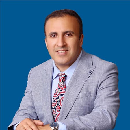
Dr. Fawwaz Al Mamoori is a consultant ophthalmologist and medical retina specialist who leads the retinal unit at Sharif Eye Centers. His training includes a me…
Hours not listedInsurance not specified7 views
Trusted medical platform in Jordan

Retinal photocoagulation, often described as laser light treatment of the retina, directs a focused beam of light onto specific areas of the retina to create tiny therapeutic spots. These seal leaking or fragile vessels, or reduce oxygen demand in areas of retina with poor blood supply so that the drive to grow abnormal new vessels subsides. It is performed in clinic under anaesthetic drops after the pupil is dilated, and you go home the same day. The commonest indications are proliferative diabetic retinopathy with its fragile bleeding vessels, focal leakage around the macula, retinal vein occlusions, thinning and tears in the peripheral retina, and retinopathy of prematurity. The technique varies with the indication: widely scattered treatment across the peripheral retina in proliferative disease, or a small number of focal spots close to a leaking point. The limits of this treatment must be stated with complete honesty. Laser is **essentially a preventive procedure that protects the sight you still have and does not give back what has been lost**. It reduces the risk of major bleeding, traction detachment and severe visual loss, but it does not repair dead tissue and does not usually improve visual acuity. It also carries an accepted cost that is explained before consent: night vision may be reduced, the peripheral field may narrow slightly, and blurring or temporary macular swelling can follow extensive sessions. That trade-off is deliberate: preserving central sight at the expense of poorly perfused peripheral retina. There is a fact more important than the procedure itself: **diabetic retinopathy is silent until advanced stages**. Your retina may already need urgent laser while your vision is still perfectly good. For that reason, **an annual eye examination for a person with diabetes is not optional**, and **controlling blood sugar, blood pressure and cholesterol slows the deterioration** and improves the result of any treatment. Laser does not cure diabetes and does not remove the possible need for intraocular injections or surgery later, and follow-up afterwards continues because the disease progresses silently. Suitability and the type of laser are decided by dilated examination and imaging, not by reading.
The pupil is dilated and the back of the eye is examined with special lenses to locate leakage, new vessels and areas of poor perfusion, while vision and eye pressure are measured. Cross-sectional retinal imaging assesses the macula, and dye angiography may be requested to map ischaemic areas precisely and guide where the laser should be applied.
It is explained that the aim is to protect vision rather than improve it, that the session may slightly change night vision or the peripheral field, and that more than one session may be required. This explanation matters, because a patient who expects sharper vision will conclude that the treatment failed and may abandon follow-up, which is the most dangerous step he could take.
Anaesthetic drops are given, you sit at the laser with your chin on the rest, and a contact lens is placed on the eye surface to direct the beam and view the periphery. For extensive sessions an injection around the eye may be used for comfort. You are asked to hold your gaze in a set direction and stay still, and this cooperation determines how precisely the treatment can be delivered.
Short pulses of light create therapeutic spots at the chosen sites, either scattered across the peripheral retina in proliferative disease or focally near a leak. You see a bright flash with each pulse and may feel pricking or mild pain that is stronger in peripheral areas, and extensive treatment is often divided over more than one appointment to reduce discomfort and swelling.
A review visit assesses whether the abnormal vessels are regressing, and further sessions may be needed to complete coverage or to treat newly affected areas. The plan is combined with intraocular injections when macular swelling is present, and the case is referred for surgery if dense bleeding or traction detachment develops. Follow-up continues lifelong, because the disease advances silently.
Bring someone to take you home, because you will not be able to drive after dilation and laser, and bring sunglasses since light sensitivity lasts for hours. Bring your most recent blood glucose reading, glycated haemoglobin result, blood pressure reading and any kidney reports, since the state of your diabetes and blood pressure shapes the course of the disease more than the session itself. Give a full list of your medicines including blood thinners and insulin, do not stop any of them on your own, and ask your doctor about the timing of your insulin dose and meal before the session if you have diabetes. Tell the doctor about any previous imaging, laser, injections or eye surgery, and about new symptoms such as flashes, floaters or a shadow in your field of vision. Eat normally and do not attend fasting unless you have been asked to.
Expect blurred vision, light sensitivity and mild headache or ache around the eye for hours up to a day or two; wear sunglasses and do not drive during this period. Use any prescribed drops, do not rub the eye, and avoid strenuous exertion and heavy lifting for the period your doctor specifies, especially if you have fragile vessels at risk of bleeding. **Seek same-day review for a sudden drop in vision, a curtain or dark shadow blocking part of your field, sudden flashes of light or a large sudden increase in floaters, or severe eye pain with redness, as these can indicate bleeding, retinal detachment or raised eye pressure.** **Keep to your periodic examinations even if your vision is excellent, because diabetic retinopathy is silent until advanced stages.** Continue working with your doctor on blood sugar, blood pressure and cholesterol control, as it is half the treatment.
Dilation and examination usually take 30 to 60 minutes before the session. The laser itself typically takes 10 to 30 minutes depending on the area treated, and extensive treatment may be divided into two or more sessions separated by days to weeks. You go home the same day, blurring lasts from hours up to two days, and a review visit is scheduled after some weeks to assess the response.
Dr. Fawwaz Al Mamoori is a consultant ophthalmologist and medical retina specialist who leads the retinal unit at Sharif Eye Centers. His training includes a me…
There are currently 1 doctor profiles linked to Retinal Photocoagulation on ClinicsJo. Review the listed services and contact the practice to confirm availability with the doctor and branch you choose.
Start with an available doctor's profile and use the contact or booking options shown there. Confirm the service, practice location and appointment time with the clinic. If this list is empty, broaden your search using the directory links on this page.
Ask the practice for the current price and what it includes. A consultation fee shown on a profile is not necessarily the price of a procedure, tests or follow-up. Confirm any additional charges and insurance arrangements before your visit.