
Amman Eye Clinic
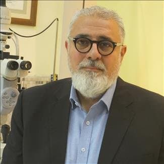
Dr. Mohammad Abu Samak is a consultant ophthalmologist with vitreoretinal fellowship training at Henry Ford Hospital in the United States. His clinical work cov…
Hours not listedInsurance not specified5 views
Trusted medical platform in Jordan

Retinal detachment surgery aims to reattach the light-sensitive layer at the back of the eye to the nourishing layer beneath it, and to seal the tear that allowed fluid to seep behind it. The technique is chosen according to the location of the tear, the extent of the detachment and the state of the vitreous gel: it may involve injecting a gas bubble into the eye combined with freezing or laser treatment of the tear, placing a silicone band around the outside of the globe, or removing the vitreous from inside the eye and filling it with gas or silicone oil that presses the retina back into place until it heals. Retinal detachment is a sight-threatening emergency. Sudden flashes of light, a dense new cluster of floaters, or a curtain or shadow blocking part of the field of vision all call for a same-day eye examination. The retina is nerve tissue that depends on the layer directly beneath it for its blood supply, and every day it stays detached means sensory cells are lost permanently, so delay costs vision that surgery cannot give back even when reattachment later succeeds. The limits of the operation matter just as much. Surgery restores the retina's position and halts further loss, but it does not promise a return to previous sharpness of vision, particularly when the detachment has reached the central macula or when a long time has passed before treatment. It does not correct short-sightedness or remove the need for glasses, it does not protect the fellow eye from a new tear, and some patients need more than one operation if scar tissue pulls the retina again. Cataract also tends to progress faster after vitrectomy in older adults. Eligibility and the choice of technique are decided by clinical examination, not by reading. The pupil is dilated and the whole retina is mapped, with macular imaging and ultrasound added when blood or opacity blocks the view inside. Tell your doctor about high myopia, previous cataract surgery or eye trauma, detachment in the other eye, or diabetes, because these change both the plan and its timing.
The pupil is dilated and the entire retina is examined to determine how many tears there are, where they sit, and whether fluid has reached the centre of vision, with macular imaging and ultrasound added when blood inside the eye blocks the view. This map drives both the choice of technique and how urgently surgery is needed.
Most cases are done under local anaesthesia given around the eye with light sedation, while general anaesthesia is preferred for children, long operations or severe anxiety. The skin is then disinfected, the face draped and the lids held open, and eye pressure is checked before the surgeon starts.
Fluid trapped behind the retina is drained, and the vitreous gel plus any membrane pulling on it is removed through three tiny ports in the wall of the eye; alternatively a silicone band is tightened around the globe to bring the wall closer to the retina in peripheral detachments. Intraocular illumination and high-magnification lenses are used throughout.
The edges of the tear are sealed with internal laser or external freezing so a permanent adhesion forms, and the eye is then filled with a gas bubble that the body absorbs over weeks, or with silicone oil that is removed at a later operation. The choice depends on the pattern of detachment and on how well you can keep the required head position.
You are given precise instructions to hold your head in a set position for a number of hours each day so the bubble presses on the tear, with review the next day and then weekly at first to check eye pressure, confirm reattachment and adjust drops. Any drop in vision between visits needs an urgent check rather than waiting for the appointment.
Tell your doctor about every medicine you take, especially blood thinners, antiplatelet drugs, prostate medicines and any pressure-lowering eye drops, and never stop a medicine on your own. Mention allergies to drugs, dressings or antiseptics, along with any heart disease, diabetes or high blood pressure, and report your recent blood sugar control if you are diabetic. Follow the fasting time you are given before the appointment, shower and clean your face, and use no make-up, contact lenses or perfume around the eye. Arrange someone to drive you both ways and to help you at home for a few days, because vision will be blurred and driving is not allowed. Tell the team about any planned air travel, since a gas bubble in the eye rules out flying until it has cleared. Bring previous reports, scans and your glasses, wear loose clothing, and do not rub the eye no matter how many floaters or flashes you notice.
Use your drops exactly on schedule and in the order prescribed, leaving a few minutes between each one, and never let the bottle tip touch the eye. Keep to the head position you were given, wear the protective shield at night during the first days, and avoid bending your head down, lifting heavy weights, straining exercise, swimming, steam and dusty places until you are cleared. Expect blurred vision and a moving horizontal line while the gas bubble is present, with gradual improvement over weeks to months, so do not judge the outcome early. Seek care immediately if you develop severe pain that painkillers do not relieve, a sudden loss of vision or the return of a curtain, increasing redness with pus-like discharge, nausea and vomiting with pain around the eye, or fever, because these can signal raised eye pressure, infection inside the eye or a new detachment. Do not fly until your surgeon confirms the gas has fully absorbed.
Surgery usually takes 45 minutes to two hours depending on the type of detachment and the technique used, with extra time for preparation and recovery. Most patients go home the same day after a few hours, though an overnight stay is sometimes advised, while the required head positioning may continue for several days up to about two weeks depending on the tamponade used.
Dr. Mohammad Abu Samak is a consultant ophthalmologist with vitreoretinal fellowship training at Henry Ford Hospital in the United States. His clinical work cov…
There are currently 1 doctor profiles linked to Detached Retina Surgery on ClinicsJo. Review the listed services and contact the practice to confirm availability with the doctor and branch you choose.
Start with an available doctor's profile and use the contact or booking options shown there. Confirm the service, practice location and appointment time with the clinic. If this list is empty, broaden your search using the directory links on this page.
Ask the practice for the current price and what it includes. A consultation fee shown on a profile is not necessarily the price of a procedure, tests or follow-up. Confirm any additional charges and insurance arrangements before your visit.